


29 Oca
Take Aorta Seriously! What is Aorta?
While Oya Aydoğan lost her life as a result of rupture of the aortic vessel, the question of what is Aort came to the agenda! So what is this aorta? DOKUZUZ Eylül University Faculty of Medicine faculty member Cardiovascular Surgeon Prof. Dr. Öztekin Oto said, 'Take the aorta seriously and do not neglect it'.
While Oya Aydoğan lost her life as a result of rupture of the aortic vessel, the question of what is aorta came to the agenda! So what is this aorta? Dokuz Eylül University Faculty of Medicine faculty member Cardiovascular Surgeon Prof.Dr. Öztekin said, 'Take the aorta seriously and do not neglect it'.
Prof. Dr. Oto said that aortic rupture (dissection) occurs as a result of rupture of the aorta, the main vessel coming out of the heart, as a result of high blood pressure and arteriosclerosis and that this is a preventable disease. Prof.Dr. Öztekin Oto said:
"When the inner layer of the vessel ruptures, pressurised blood enters between the three layers of the vessel wall. This second blood pathway collapses the main flow path of the vessel and disrupts the blood circulation of vital organs such as the brain, arms and legs. The most common causes of all these are high blood pressure and arteriosclerosis. If the blood pressure, which can also be defined as the pressure created by the blood in the artery on the walls, is too high, it can peak at a moment with straining, coughing or straining and cause a rupture (dissection) in the vessel wall. Therefore, patients with high blood pressure should take medication to keep their blood pressure below 135/85 mmHg. In addition, patients with high blood pressure should undergo echocardiography at appropriate intervals to check whether there is enlargement of the left ventricle of the heart or the aorta. In this way, the risk of aortic rupture can be reduced and even prevented."
Stating that severe pain, loss of consciousness and sudden paralysis may occur in aortic dissection, Prof. Dr. Öztekin Oto continued his speech as follows:
"In this case, the patient can be saved if a rapid echocardiography and computed tomography are performed and an intervention is performed immediately by an experienced cardiovascular surgeon. The risk of death increases by 1 per cent for every hour of delay between diagnosis and surgery. Therefore, the 'door-table time' between entering the hospital door and being placed on the operating table should be kept very short. While in the past we used to stop the heart and cool the patient down a lot during the operation, now we cool down less, use special brain protection methods, replace the ruptured aorta and connect the vessels to the brain and arms to this new vessel. Thus, the patient's blood vessel can be replaced while preserving the circulation of vital organs."
WHAT IS AORTA?
The aorta is the largest artery in the human body. It originates in the left ventricle and runs down the abdomen and then branches into smaller vessels. The aorta delivers oxygenated blood to all body parts through the systemic circulation.
Direction of the aorta
Course of the aorta (anterior view), starting posterior to the main pulmonary artery, but then anterior to the right pulmonary arteries, the trachea and the esophagus, but then turning posteriorly to course dorsally to these structures.
The aorta is usually divided into five segments or parts.
Ascending aorta: The segment between the aortic arch and the heart.
Aortic arch: The apex that looks like an inverted U.
Descending aorta: A segment of the aortic arch that divides into the iliac arteries.
Thoracic aorta: Half of the descending aorta above the diaphragm.
Abdominal aorta: Half of the descending aorta below the diaphragm.
Embryological Development
In mammalian and avian embryological development, pharyngeal arch (aortic arch) arteries have similar patterns to normal arteries. The fourth aortic arch vessel survives in these vertebrae as the aortic arch. The third aortic arch vessel continues as the root of the brachiocephalic artery or internal carotid artery. The sixth arch is joined by the pulmonary arteries. The smooth muscle of the great artery and the cell community in the aortapulmonary septum are divided into the aorta and pulmonary arteries by the cardiac neural crest. The involvement of smooth muscle of this neural crest in the great artery is rare, with many smooth muscles derived from mesoderm. In fact, smooth muscle is derived from mesoderm in the abdominal aorta and only the coronary arteries, which arise above the semilunar valves, have a mesodermal origin of smooth muscle. The lack of division of the aorticopulmonary septum into large vessels causes persistent truncus arteriosus.
Click here to go to the news source.
Related Articles

Tiny from Adana Found Life in Izmir
Melek Özcan, 2.5-year-old Melek Özcan, who was waiting for an organ at Dokuz Eylül University Hospit..
Read More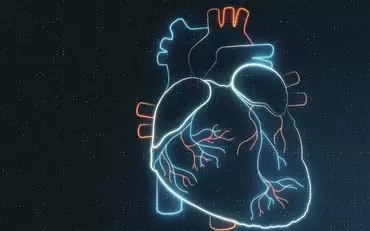
They clung to life with artificial heart
In Izmir, 3-year-old Melek Özcan, who was given Turkey's smallest artificial heart to hold on to lif..
Read More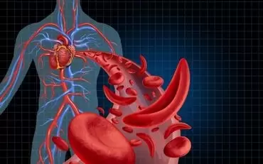
President of the Surgical Association
At a press conference held at the Dean's Office of DEU Faculty of Medicine, Prof. Dr. Oto stated tha..
Read More